


Image Reconstruction
Principle of Image Reconstruction
Image reconstruction is a fundamental technique that unlocks valuable insights from raw data. This process involves multiple critical components working in harmony to transform collected signals into a meaningful visual representation.
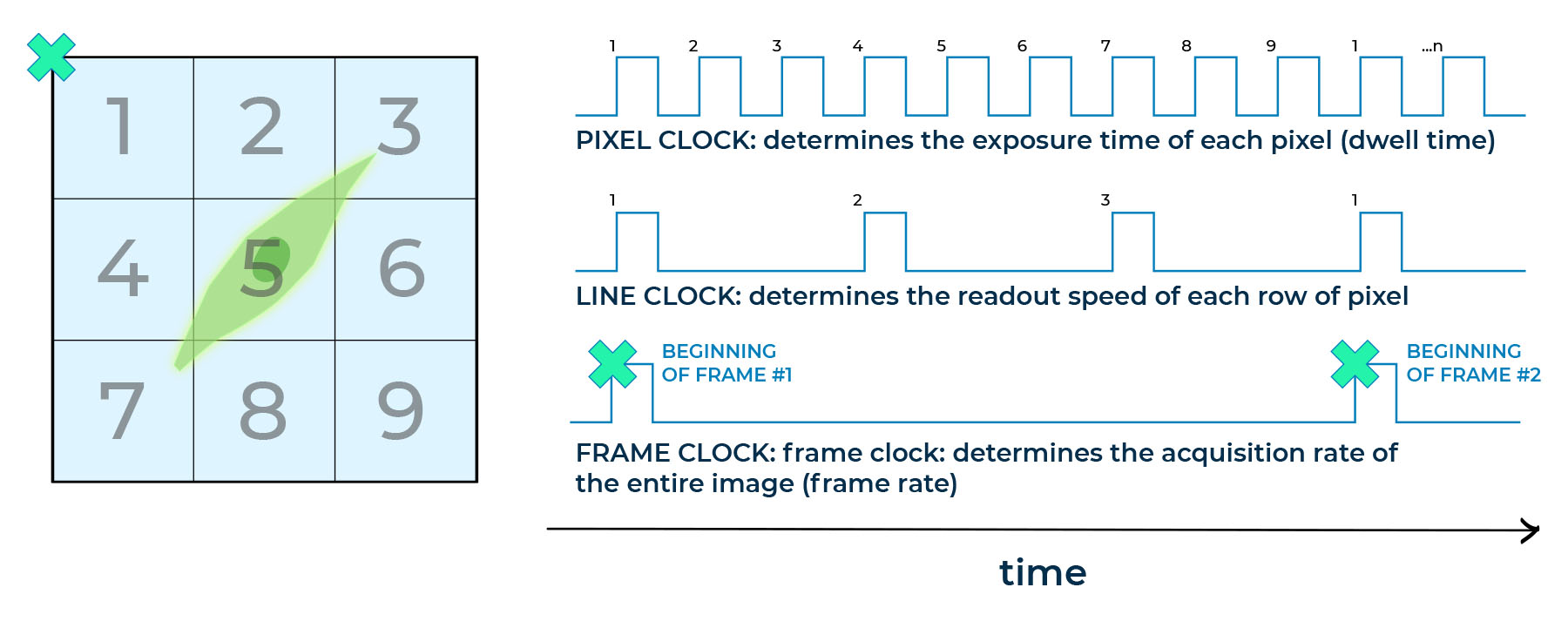
At the heart of this process lies the scanning system, comprising scanning mirrors and a focusing lens. These components delicately control the movement of a laser beam across the sample, pixel by pixel. Alternatively, a motorized stage can also achieve pixel-by-pixel scanning, controlling sample motion while the laser beam remains fixed. This careful control is essential for the accurate spatial information collection through integrated pixel, line, and frame/scan clocks.
Scanning System
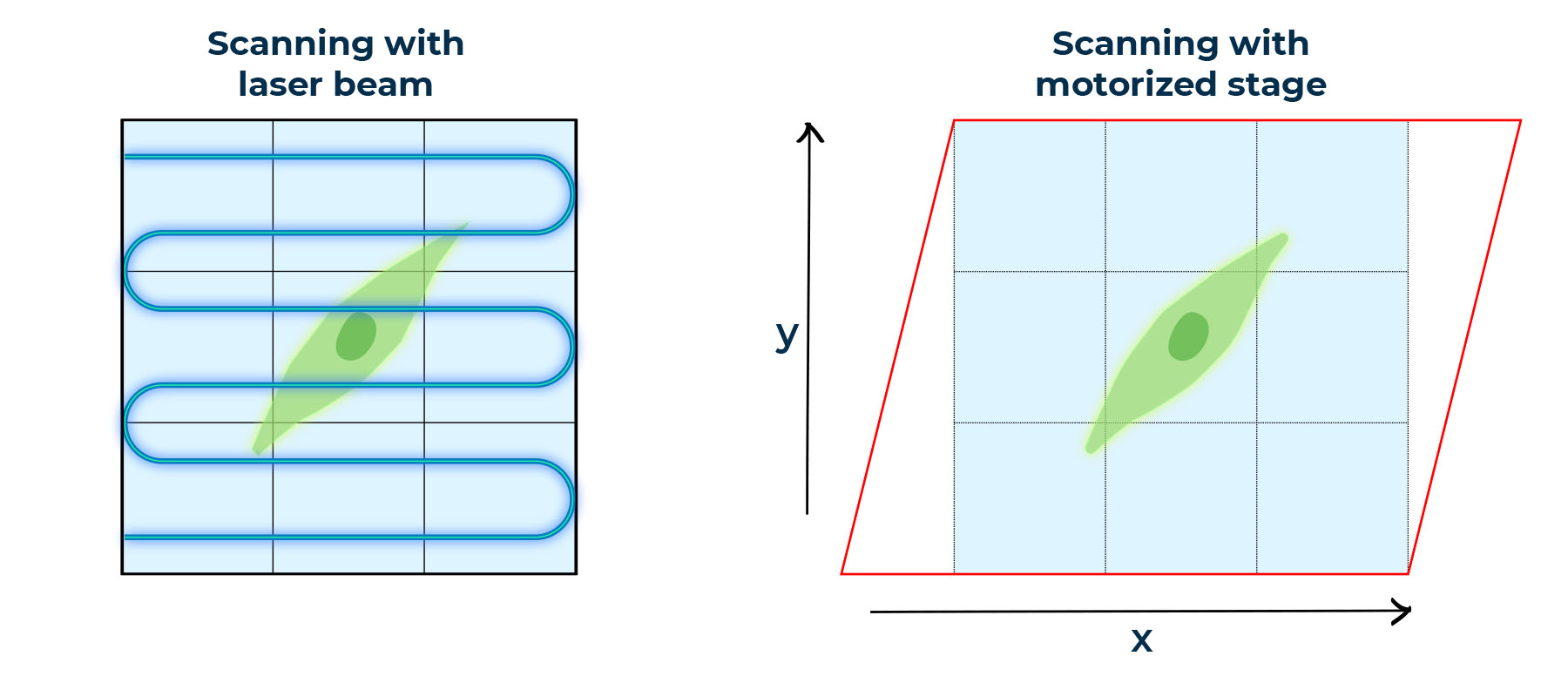
An exaggerated cell sample scanned using two pixel-to-pixel scanning system:
scanning with laser beam: the blue path indicates the scanning process;
scanning with motorized stage: red parallelogram suggests the motorized stage.
Images are made out of 9 pixels (3×3 pixels image).
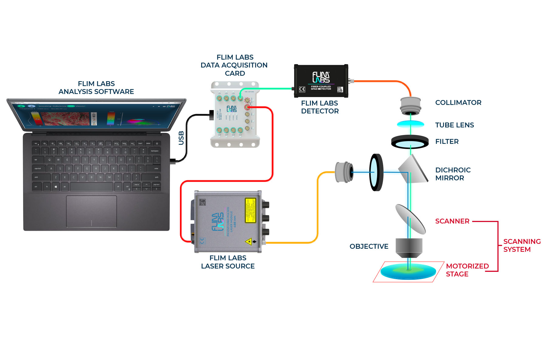
When the laser beam interacts with the sample, it triggers the emission of fluorescent light. Detectors capture this emitted light, generating a signal that holds vital information about the sample.
Both the spatial information and the captured signal are then transmitted to the FLIM Data Acquisition Card. In essence, the data acquisition card plays a pivotal role, collaborating with other components to ensure precise image reconstruction.
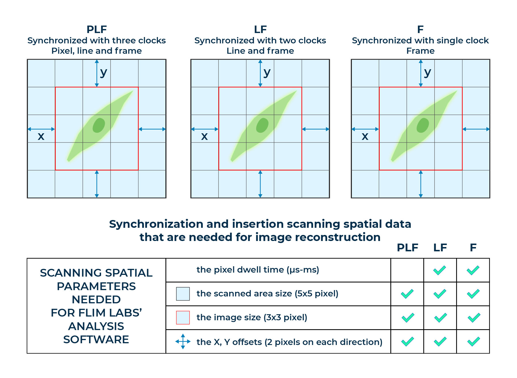
Once all the data is recorded in data acquisition card and transferred to the software, the insertion of spatial parameters, including scanned area size, actual image dimensions, and X, Y offsets in pixel units, culminates in image reconstruction. This visual representation unveils the distribution of fluorophores within the sample, allowing researchers to gain valuable insights.
Essential Elements for Image Reconstruction: (1) Laser-induced fluorescent light emission from sample; (2) Detector capturing the light signal and transmitting it to an acquisition card; (3) Synchronization of spatial scanning data with the acquisition card; (4) Insertion of spatial parameters, enabling software to unveil the distribution of fluorescent molecules within the sample.
FLIM LABS’ FLIM Data Acquisition Card Empowers Image Reconstruction
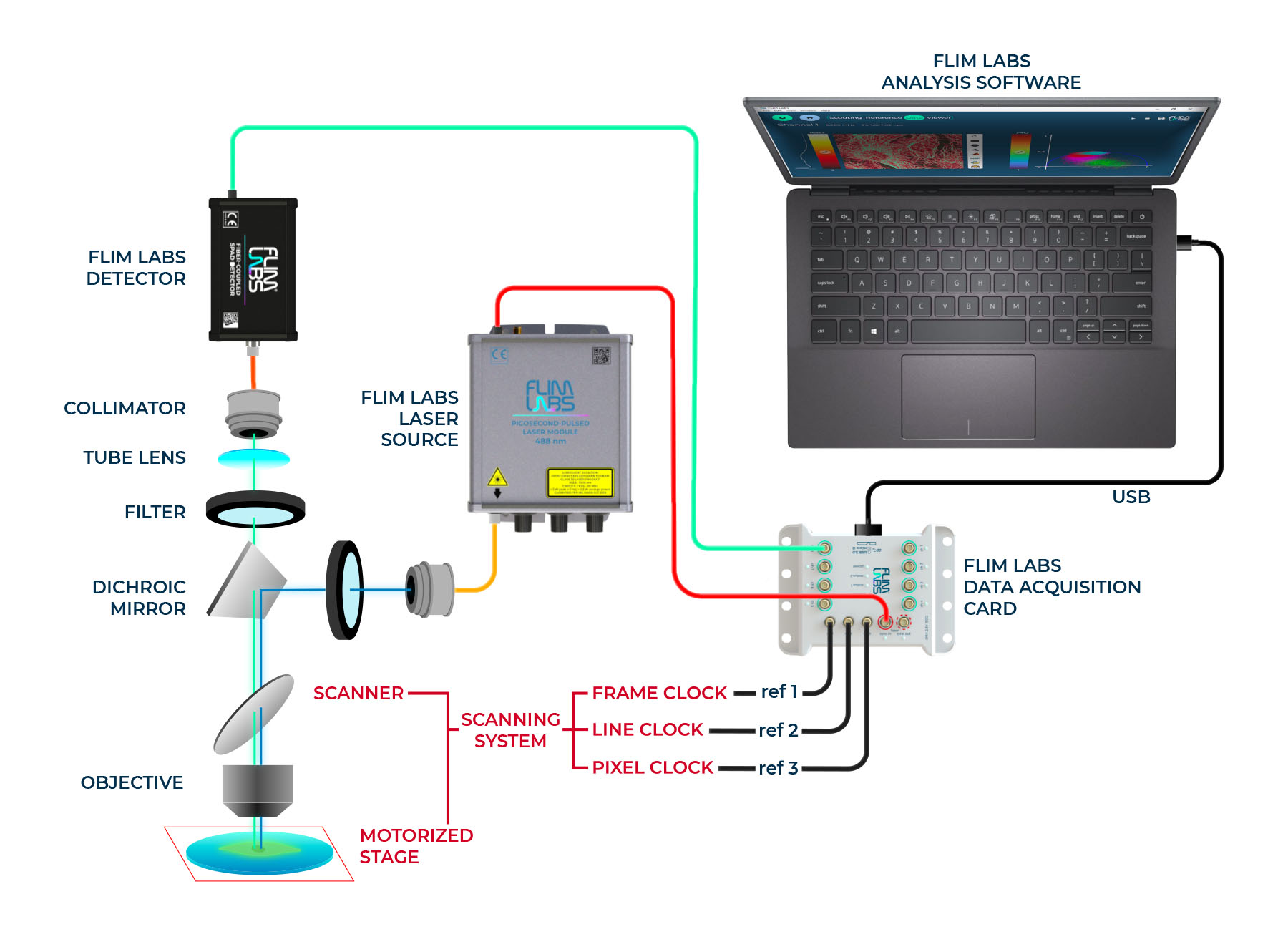
FLIM LABS’ FLIM Data Acquisition Card serves as a bridge uniting the scanning system, detectors, and data processing software. This diligent card receives all the necessary data for accurate image reconstruction using different input channels for synchronization. Three specialized input channels enable precise spatial information acquisition:
- Ref 1: Frame/scan clock synchronization
- Ref 2: Line clock synchronization
- Ref 3: Pixel clock synchronization
FLIM LABS’ Data Acquisition Card provides ref 1 channel to synchronize with frame clock, ref 2 channel to synchronize with line clock and ref 3 channel to synchronize with pixel clock.
The card’s precision in synchronization guarantees faithful data transformation into a reconstructed image.
Even in cases where only a single synchronization signal is available, our accompanying software seamlessly overcomes challenges. Leveraging information from the frame/scan clock alone or together with the line clock, the software adapts. By incorporating the pixel dwell time with other spatial parameters (scanned area size, actual image dimensions, and X, Y offsets in pixel units), the software tailors itself to various experimental setups, making image reconstruction accessible to all researchers.