FLIM-Phasors


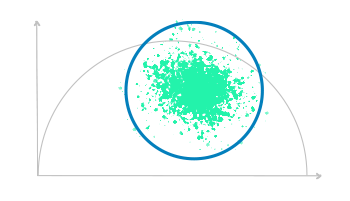
The information contained in a FLIM image can be easily handled when the phasor analysis approach is introduced. This method allows a graphical representation (phasor plot) of the lifetime distribution. Indeed, the interpretation of the FLIM phasor plot is straightforward and the combination of FLIM and phasors enables the researchers to easily separate different lifetime populations within the same FLIM image [1].
The phasor plot figure illustrates an exaggerated FLIM phasor analysis example with three circlets, each representing a different lifetime value (T1 in red, T2 in green, T3 in blue). The circlets vary in size for visual clarity, showing T1 as the smallest, followed by T2 and T3.
Different fluorophores with distinct lifetimes can be discriminated because every species is associated with a precise phasor (the so-called “fingerprint”). Using the geometrical working principle at the basis of the phasor-plot, different lifetime values (a cloud of points) are distributed over a semicircle where the “longest” lifetime are on the left side, while the “shortest” ones on the right side of the plot.
Moreover, if the fingerprint cloud falls precisely on the semicircle, it means that the fluorescence signal can be described by a single exponential decay, while if the cloud falls within the semicircle area, the lifetime is a superposition of different values. Therefore, the FLIM-phasor approach is extremely straightforward for the interpretation of fluorescence lifetime fingerprints in the sample under investigation [2].
In our software FLIM STUDIO, the flim-phasor approach, combined with tailored circlets, helps researchers extract meaningful insights about fluorescence lifetime analysis in specific regions. See the demo video for a quick tutorial.
[1] Digman MA, Caiolfa VR, Zamai M, Gratton E, The phasor approach to fluorescence lifetime imaging analysis. Biophys J. 2008 Jan 15;94(2):L14-6.
doi: 10.1529/biophysj.107.120154
Epub 2007 Nov 2. PMID: 17981902; PMCID: PMC2157251
[2] Digmann MA, Gratton E, The phasor approach to fluorescence lifetime imaging: Exploiting phasor linear properties (2012)
https://escholarship.org/uc/item/5g279175
[3] Vallmitjana A, Torrado B, Gratton E, Phasor-based image segmentation: machine learning clustering techniques. Biomed Opt Express.
2021 May 17;12(6):3410-3422.
doi: 10.1364/BOE.422766
PMID: 34221668; PMCID: PMC8221971