Home › Methods › FLIM-FRET

Methods

Förster Resonance Energy Transfer with FLIM

A short introduction to FRET and how fluorescence lifetime measures it.

FRET reports whether two molecules are within a few nanometres of each other, well below the ~250 nm diffraction limit of light microscopy.

FLIM-FRET reads it from the donor’s fluorescence lifetime, which shortens when an acceptor is close, giving a measurement that is largely independent of probe concentration.

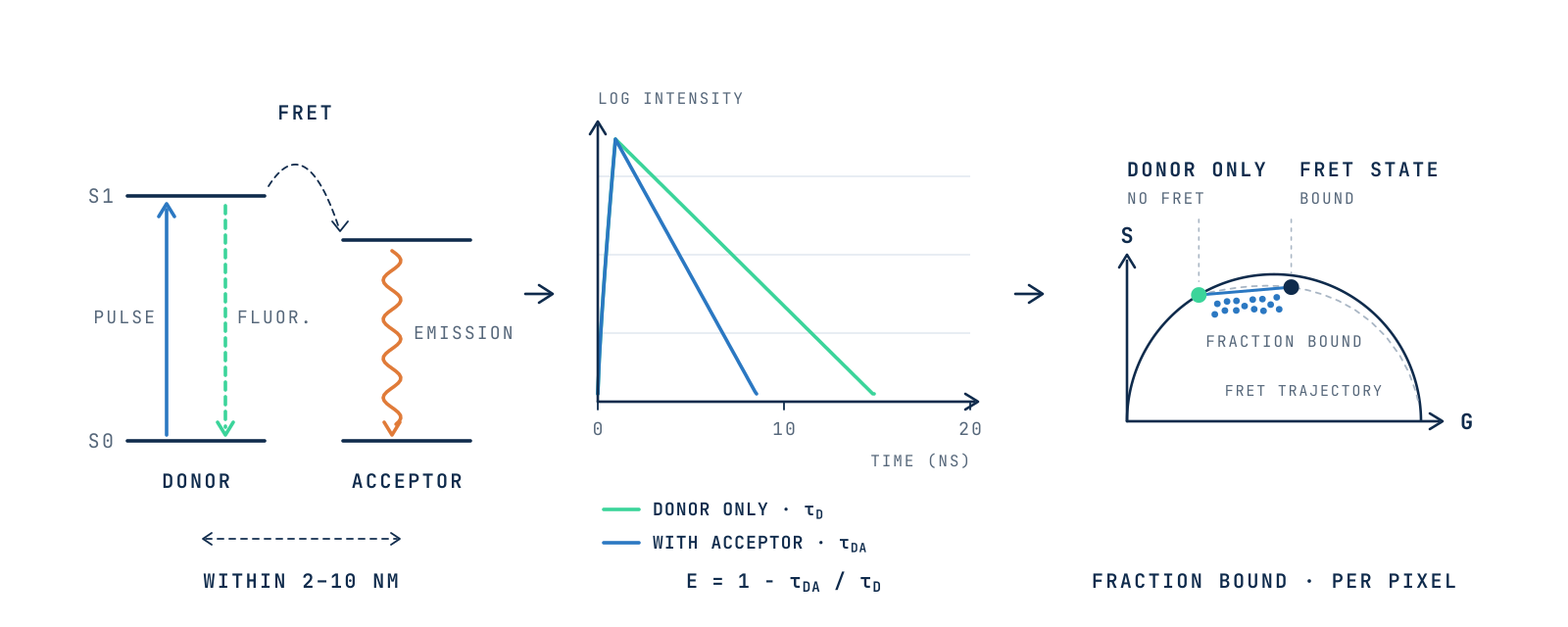
FLIM-FRET: from energy transfer to the phasor plot

When an acceptor is within a few nanometres, the excited donor transfers part of its energy to it, so the donor lifetime gets shorter. On the phasor plot, each pixel then lies between the donor-only and FRET states, giving the fraction of interacting molecules. Schematic.

01 FRET

What is FRET?

Förster Resonance Energy Transfer (FRET) is the transfer of energy from an excited donor fluorophore to a nearby acceptor. After absorbing a photon, the donor can return to its ground state through fluorescence or other competing pathways. When an acceptor is close enough, an additional pathway opens: the donor passes its energy directly to the acceptor through dipole–dipole coupling.

The transfer is non-radiative: no photon is emitted by the donor and reabsorbed. The donor returns to the ground state, while the acceptor becomes excited and may emit its own fluorescence.

FRET energy transfer

Figure 1 · The excited donor can fluoresce or transfer its energy to the acceptor, which then emits. The transfer efficiency falls steeply with distance around the Förster radius R0. Schematic.

02 Conditions

When does FRET occur?

FRET is efficient only when three conditions are met at the same time.

01

Proximity

Donor and acceptor are about 2–10 nm apart; beyond 10 nm FRET becomes negligible. The transfer rate scales with the inverse sixth power of the distance, so efficiency changes steeply around the Förster radius (R0, the distance at which E = 50%).

02

Spectral overlap

The donor’s emission spectrum overlaps the acceptor’s absorption spectrum.

03

Orientation

The transition dipoles of the two fluorophores are favourably aligned. When both rotate quickly during the donor lifetime, orientation averages out (κ2 = 2/3).

03 Lifetime

Why measure FRET by lifetime?

FRET opens an extra route out of the donor’s excited state, so the donor lifetime gets shorter. FLIM-FRET measures this shortening directly, and the FRET efficiency follows from it: E = 1 − τDA/τD.

01

Largely independent of concentration

Uneven donor expression does not bias the lifetime, although very dim samples and the acceptor-to-donor ratio still need care.

02

Donor only

No spectral bleed-through correction is needed, and even dark acceptors can be used.

03

Non-destructive

No acceptor photobleaching, so the same living cell can be measured repeatedly.

Donor decay with and without acceptor

Figure 2 · In the presence of the acceptor, the donor decays faster. The lifetime change gives the FRET efficiency.

04 Analysis

How are FLIM-FRET data analyzed?

Donor decays can be fitted with exponential models, or analyzed without fitting using phasor analysis. In the phasor plot, FRET moves the donor along a calculated trajectory away from the donor-only position. Once background, autofluorescence and non-interacting donors are taken into account, each pixel’s position gives the FRET efficiency and the fraction of interacting molecules.

FRET in the phasor plot

Figure 3 · Phasor analysis separates free and interacting donors in each pixel without curve fitting.

Controls

Reliable measurements need controls, measured under the same conditions as the sample: a donor-only sample to set τD, the donor with a non-interacting or free acceptor as negative control, and ideally a donor–acceptor fusion as positive control. In tissues and plants, an unlabeled sample also records the autofluorescence background.

05 Applications

What can FLIM-FRET reveal?

From interacting proteins to living-cell signalling, lifetime turns molecular proximity into a quantitative readout.

01

Protein–protein interactions and complexes

Whether molecules bind, what fraction is bound, and where, from dimers and receptor complexes to chromatin compaction at the nucleosome level.

See the Molecular interactions application

02

Conformational changes

Shape changes within a single molecule, such as a biosensor switching between states or G-protein rearrangement on activation.

03

Biochemical activity

Biosensors reporting GTPase, kinase and second-messenger activity in living cells over time, now fast enough to screen signalling kinetics across hundreds of cells.

Further reading

References

1Digman MA, Caiolfa VR, Zamai M, Gratton E. The phasor approach to fluorescence lifetime imaging analysis. Biophysical Journal. 2008;94(2):L14–L16. doi:10.1529/biophysj.107.120154

2Hinde E, Digman MA, Welch C, Hahn KM, Gratton E. Biosensor Förster resonance energy transfer detection by the phasor approach to fluorescence lifetime imaging microscopy. Microscopy Research and Technique. 2012;75(3):271–281. doi:10.1002/jemt.21054

3Eljebbawi A, Dolata A, Strotmann VI, Weidtkamp-Peters S, Stahl Y. From principles to practice: a comprehensive guide to FRET-FLIM in plants. Methods in Microscopy. 2025;2(1):33–43. doi:10.1515/mim-2024-0019

4Lou J, Scipioni L, Wright BK, et al. Phasor histone FLIM-FRET microscopy quantifies spatiotemporal rearrangement of chromatin architecture during the DNA damage response. PNAS. 2019;116(15):7323–7332. doi:10.1073/pnas.1814965116

5Coucke Q, Parveen N, Solís Fernández G, et al. Particle-based phasor-FLIM-FRET resolves protein-protein interactions inside single viral particles. Biophysical Reports. 2023;3:100122. doi:10.1016/j.bpr.2023.100122

6Szabó Á, Szöllősi J, Nagy P. Principles of resonance energy transfer. Current Protocols. 2022;2:e625. doi:10.1002/cpz1.625

7Harkes R, Kukk O, Mukherjee S, Klarenbeek J, van den Broek B, Jalink K. Dynamic FRET-FLIM based screening of signal transduction pathways. Scientific Reports. 2021;11:20711. doi:10.1038/s41598-021-00098-9

From principle to measurement

FLIM-FRET on your microscope

FLIM LABS upgrade kits add TCSPC lifetime acquisition to compatible confocal and multiphoton microscopes, with phasor analysis in FLIM IMAGER.